
The Patient
A 49-year-old female equestrian. Equestrian sport carries a specific and serious risk profile for the cervical spine — repeated impact loading through the saddle, the constant postural demands of riding, and the ever-present possibility of falls. Her history reflected that reality. The structural changes on her X-rays were consistent with years of trauma-induced subluxation that had been left uncorrected.
What showed up: loss of cervical curvature, degenerative disc thinning — those dark spaces between the vertebrae that indicate the discs have lost height under chronic abnormal loading — and changes to the bone architecture consistent with long-term structural stress. She completed a 6-month initial correction plan and has elected to continue into a follow-up 3-month plan based on the spinal and functional improvements she experienced.
What the X-Rays Showed — and How to Read Them
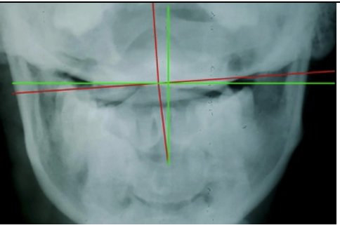
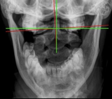
On her re-evaluation films, something stands out that requires explanation: her head tilt on the AP (front-to-back) cervical view appears different than her initial — and at first glance, it may look unfavorable. I want to address that directly, because it is one of the most important concepts in structural correction.
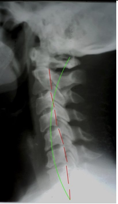
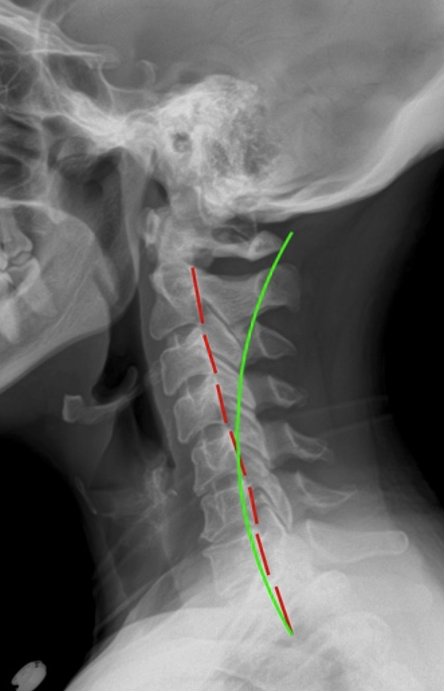
Look at the lateral cervical view. Her curve went from reversed at baseline to almost straight at re-evaluation. That is significant structural correction. A reversed cervical curve means the spine is bending the wrong direction — placing the spinal cord and nerve roots under chronic tension, loading the discs at wrong angles, and creating sustained mechanical stress on the brain stem. Moving from reversed toward straight is movement in the right direction.
When the spine undergoes that kind of structural transition, the AP view will look different. The head alignment shifts as the underlying curve reorganizes. What appears as a change in head tilt on the AP is the structural consequence of a spine in the process of correcting. Progress must be read across both views, in light of the full structural picture — not from a single measurement in isolation.
Functional Improvement: The Proof in the Saddle
The structural changes confirm what she already knew from her experience. The spinal and physiological improvements she noticed during her initial correction plan were what motivated her to continue. And those improvements have shown up where it matters most to her — in her riding.
That is not incidental. Equestrian performance depends heavily on the nervous system's ability to coordinate balance, proprioception, and the precise neuromuscular communication between rider and horse. When cervical subluxation is creating interference in those pathways, the rider compensates — often without knowing it. As the subluxation is corrected and nerve flow is restored, that compensation pattern releases. The body moves more freely and more accurately. The horse responds.
Outcomes After Initial Correction Plan
Lateral cervical curve moved from reversed toward normal lordotic arc.
Brain stem stress reduced; nerve flow between brain and body progressively restoring.
Disc degeneration and bone changes stabilizing as mechanical stress is removed.
Measurable improvement in spinal alignment across both AP and lateral views.
Functional improvement confirmed in equestrian riding performance.
Continuing into follow-up 3-month correction plan for further structural restoration.
Why This Pattern Matters
Equestrian sport creates a specific and serious cervical risk profile that accumulates over a career. Each ride subjects the cervical spine to impact loading transmitted through the saddle, constant postural demand to stabilize the head while the horse moves beneath, and the ever-present possibility of falls that load the neck in angles it was not designed to absorb repeatedly. The result — when the structural consequence goes uncorrected — is a cervical curve that reverses under that sustained mechanical pressure. Instead of the normal lordotic arc distributing load evenly, the reversed curve concentrates it. Disc degeneration follows. Nerve compromise follows. The rider compensates without knowing it.
What makes this case instructive beyond the structural findings is the functional confirmation: her improvement showed up in her riding. That is not incidental. Equestrian performance is neurological. Balance, proprioception, the fine neuromuscular communication between rider and horse — all of it depends on a nervous system that can signal clearly and receive clearly. Cervical subluxation at the level of the brain stem compromises exactly those pathways. Correcting it restores them. The horse notices. The rider notices. The X-ray confirms what both already knew.
What to Look For
Riders with cervical subluxation typically present with neck stiffness or tension that intensifies after rides, reduced rotation to one side that limits their ability to look freely in both directions, and headaches that appear predictably following riding sessions. These are not the expected costs of the sport. They are structural signals. So is any performance plateau or regression — particularly in balance, timing, or feel — that does not respond to better technique, better equipment, or additional training hours.
Any rider with a history of falls — even minor ones over the years — carries a cervical trauma history. The damage does not have to be dramatic to be structurally significant. A reversed cervical curve takes years to develop. The falls that contributed to it are often forgotten long before the structural consequence becomes undeniable. If you ride and you have never had a structural cervical assessment, the X-ray will show you exactly where your spine has absorbed the history of your sport. Have you ever had your spine checked for subluxation?
Has Trauma Changed Your Spine's Structure?
Physical trauma — from sports, accidents, or years of repetitive loading — leaves structural evidence in the spine long after the acute event. A structural assessment at Rochet Family Chiropractic reveals exactly what that evidence shows and whether corrective care can restore what was lost. Serving Royal Palm Beach, Wellington, and the greater West Palm Beach area.
Schedule a Structural AssessmentOr call us at (561) 795-3156
To learn more about how cervical subluxation affects structural integrity and nervous system function, visit our cervical subluxation resource page.