What He Came In For
This patient had been under care with us and was returning for a follow-up spotlight — we featured him months earlier after his initial corrective plan. His original complaint was low back pain. That is what brought him through the door. Low back pain is one of the most common reasons people seek chiropractic care in Royal Palm Beach, and in most cases the assumption is that the low back is where the work needs to happen. In his case, the X-rays told a different story from the very beginning.
When we examined his spine, his greatest vertebral subluxation was not in his lumbar spine. It was in his Atlas (C1) and Axis (C2) — the two uppermost vertebrae of the cervical spine, positioned directly at the base of his skull. This is more common than most people realize. The spine is one continuous structure, and the area of discomfort is frequently not the area of greatest subluxation.
What the X-Rays Revealed
Clinical Findings — Initial Examination
Primary subluxation: Atlas (C1) and Axis (C2) — upper cervical spine
Presenting complaint: Low back pain
X-ray type: AP upper cervical
Finding: Significant C1 and C2 displacement confirmed on AP film
Adjustment distribution: Over 80% of all corrective adjustments delivered to Atlas (C1)
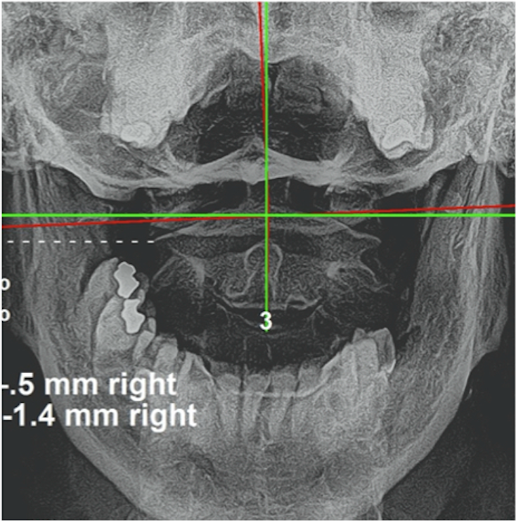
The AP upper cervical X-ray showed measurable displacement of C1 and C2 relative to the skull base. The Atlas is a ring-shaped vertebra with no disc above it — it articulates directly with the occipital bone of the skull. Its position has a direct mechanical relationship to the brainstem, which passes through it. Subluxation of C1 and C2 does not stay local. The neurological effects travel.
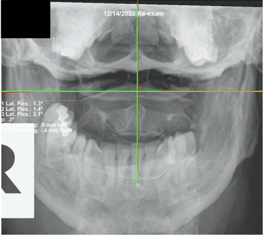
Atlas (C1) & Axis (C2) AP — Before & After
How We Approached His Care
Because the Atlas and Axis were the primary subluxations, the vast majority of his corrective adjustments were delivered to C1. That is not instinct — that is what the X-ray directs. We do not guess where to adjust. The film tells us. His low back received attention as a secondary site, but the structural correction priority was always upper cervical.
C1 and C2 have a direct influence on the brainstem, and through the brainstem, on every organ system and function in the body. When the Atlas is subluxated, the body compensates throughout the spinal column — creating secondary patterns that show up as discomfort in areas the patient thinks of as “the problem.” Correcting the primary subluxation allows the body, through Innate Intelligence, to begin unwinding those compensatory patterns on its own.
What Happened — and Why He Kept Going
After completing his initial six-month corrective plan, this patient elected to continue with an additional three-month phase. That decision tells you more than the X-rays alone. His quality of life had improved to a degree that made the answer obvious to him. Sleep had improved. Energy had improved. The way he managed daily stress had improved. These are not outcomes we promise — they are what the body does when vertebral subluxation is corrected and the brainstem has the structural room it was designed to have.
The re-evaluation X-ray taken December 14, 2020 confirmed what he was experiencing: proper alignment of C1 and C2 had been achieved. The structural work reflected the functional improvements. He came in for low back pain. He left with a spine that was doing what it was created to do.
I am not the healer. I correct the subluxation. God designed the body to heal itself when the interference is removed — and when you are subluxation free, the body expresses life the way it was created to.
Why This Pattern Matters
The atlas case is one of the most instructive patterns in subluxation-based chiropractic. Most people — and most practitioners — default to treating where the pain is. When a patient comes in with low back pain, the assumption is that something is wrong in the low back. Sometimes that's true. But the spine is one continuous mechanical and neurological unit. The atlas (C1), at the very top of the cervical spine, is the keystone. Its position influences the entire column below it.
When C1 is subluxated, the body compensates. The pelvis tips. The lumbar spine shifts to balance what's happening above it. The thoracic spine follows. What the patient experiences as low back pain is often the bottom of a compensatory chain that started at the top. Correct the atlas, and the compensations below have a chance to resolve — because the primary interference has been removed.
Innate Intelligence is always working toward adaptation and balance. What it cannot do is correct a structural displacement on its own. Remove the primary subluxation, and the body, through Innate Intelligence, begins to restore the pattern it was designed to maintain.
The resolution timeline in this case — six months of initial correction followed by three additional months — reflects the biology of structural unwinding. The compensatory chain that formed from C1 displacement didn't develop overnight, and it does not resolve overnight. The pelvis had been off-level. The lumbar column had been loaded asymmetrically. Soft tissue had adapted to those positions. Each level of that compensation had to reorganize as the atlas correction held. This is why care plans in this work are measured in months rather than visits, and why the results continued to improve as time allowed the body to process each structural change.
The functional improvements this patient reported — sleep quality, energy, and daily stress management — are nervous system outcomes, not symptoms that were treated. The brainstem, freed from mechanical obstruction at C1, could regulate the autonomic functions it governs without the compression that had been limiting them. Those functions include sleep cycle regulation, metabolic energy production, and the body's capacity to respond to daily demand. Correcting the atlas did not target any of those functions directly. It removed the structural interference that was restricting all of them.
What to Look For
Upper cervical subluxation — particularly at C1 and C2 — produces a wide range of effects because of its proximity to the brainstem. The signs are often not what people associate with a neck problem. Disturbed sleep. Low energy that doesn't resolve with rest. Difficulty managing daily stress. Headaches that start at the base of the skull. A sense that the head isn't sitting quite straight. These are brainstem-level effects, not the signs of a sore neck.
In patients with low back complaints, look also for any of the above. The lumbar finding may still be secondary to what's happening at C1. That's why X-ray analysis — not assessment based on where it hurts — is the only reliable way to find the actual subluxation. The spine keeps no secrets from a good film.
The compensatory lumbar pattern also has specific characteristics worth recognizing: low back tension that is consistently worse on one side; a history of recurring episodes that partially resolve but never fully clear; the sense that one hip sits higher or one leg seems shorter when standing. These are the body's mechanical expression of a compensation chain that originates above the lumbar spine. When these presentations arrive without a clear lumbar history — no specific injury, no disc finding, no identifiable local cause — the upper cervical spine deserves direct evaluation.
Upper cervical subluxation driving lumbar compensation is also worth considering in any patient who has had consistent care and still finds the same mechanical pattern reasserting. If the low back adjustment has been regular, if the pain response has been partial and cyclical, and if the atlas has never been specifically evaluated on an AP cervical X-ray — the source of the pattern may be above the region being treated. The structural chain runs from C1 down. When the top is displaced, the correction below remains incomplete. Have you ever had your spine checked for subluxation?
Your Biggest Subluxation May Not Be Where You Think It Is
Most people come in focused on where they feel it. The X-ray shows us where the structural problem actually is. If you have been managing low back discomfort, neck stiffness, or any recurring issue without a structural evaluation, you owe it to yourself to find out what is actually happening in your spine. Come in for a consultation and X-ray analysis.
Schedule a Structural EvaluationOr call us at (561) 795-3156
Related: How atlas subluxation manifested as facial tics in a 10-year-old — another case where the upper cervical spine was the key.